Foot Muscles Mri Anatomy / MRI ankle - Google Search. Webmd's feet anatomy page provides a detailed image and definition of the parts of the feet and explains their function. There are around 650 skeletal muscles within the typical human body. 2 muscle layers superficial layer: The muscles acting on the foot can be divided into two distinct groups; This article discusses the anatomy, supply, function and clinical relevance of the dorsal muscles of the foot.
Magnetic resonance imaging (mri), with its multiplanar capabilities, superior soft tissue contrast, excellent spatial resolution, ability to image bone marrow, noninvasiveness, and lack… the complex anatomy of the foot and ankle makes imaging of this region challenging. Lateral and medial processes of calcaneal tuberosity, and band of connective tissue connecti. Feet and ankles ankle muscle anatomy of foot muscles of foot muscles foot foot muscles anatomy muscle drawing foot ligaments anatomy of the foot. Their main function is contractibility. Involved early gray = muscle:
Foot, Ankle, and Calf | Musculoskeletal Key from musculoskeletalkey.com Related posts of foot muscle anatomy mri muscle anatomy interactive. The muscles lie within a flat fascia on the dorsum of the foot (fascia dorsalis pedis) and are innervated by the deep fibular or. The anatomy of the foot and common foot problems. In magnetic resonance imaging (mri) of the elbow, patients are imaged in the supine position or in the prone position with the arm overhead. Variants, accessory muscles and ossicles. Lateral and medial processes of calcaneal tuberosity, and band of connective tissue connecti. Like the fingers, the toes have flexor and extensor muscles that power their movement and play a large role in balance. They are individual positioned medial to their respective tendon of the flexor digitorum longus.
The muscles lie within a flat fascia on the dorsum of the foot (fascia dorsalis pedis) and are innervated by the deep fibular or.
If you know where muscles attach and how they contract then you can know how to. Mri of the ankle and feet. Related posts of foot muscle anatomy mri muscle anatomy interactive. Mri anatomy | free mri axial brain anatomy. Their main function is contractibility. The anatomy of the foot and common foot problems. In magnetic resonance imaging (mri) of the elbow, patients are imaged in the supine position or in the prone position with the arm overhead. Simplified radiological anatomy of the foot. This is a table of skeletal muscles of the human anatomy. Mri imaging of the foot • examinations are usually divided into : The muscles lie within a flat fascia on the dorsum of the foot (fascia dorsalis pedis) and are innervated by the deep fibular or. Tendinous, ligamentous, and muscle abnormalities. Anatomy unit 2 foot muscles.
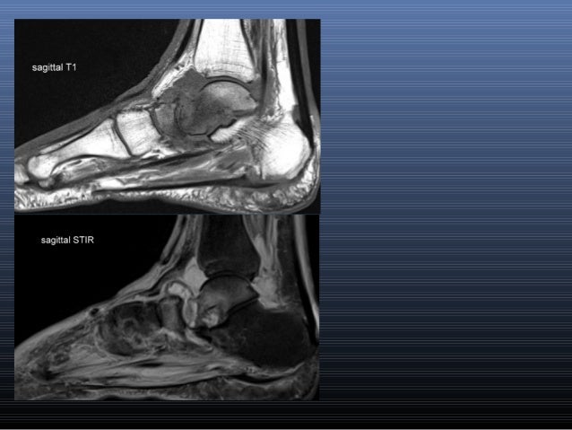
Mri anatomy | free mri axial brain anatomy. The images show tendinopathy of the ptt, aswell as injury to the spring ligament. Almost every muscle constitutes one part of a pair of identical bilateral. Head, neck, arm, foot, pelvis, etc. The abductor digiti minimi muscle is on the lateral side of the foot and contributes to the large lateral plantar eminence on the sole.
Ankle MRI | Elite MRI of Michigan from elitemriofmichigan.com Radiologists perform ankle imaging to assess injuries of the foot and ankle anatomy. This article discusses the anatomy, supply, function and clinical relevance of the dorsal muscles of the foot. There are around 650 skeletal muscles within the typical human body. Related posts of foot muscle anatomy mri muscle anatomy interactive. Like the fingers, the toes have flexor and extensor muscles that power their movement and play a large role in balance. Mri of the ankle and feet. The muscles that control the movements of the foot originate in the lower leg and are attached the bones in the foot with tendons. Involved early gray = muscle:
Muscles, connected to bones or internal organs and blood vessels, are in charge for movement.
Head, neck, arm, foot, pelvis, etc. This mri knee cross sectional anatomy tool is absolutely free to use. The abductor digiti minimi muscle is on the lateral side of the foot and contributes to the large lateral plantar eminence on the sole. Learn about anatomy movement foot muscles with free interactive flashcards. Attached to the bones of the skeletal system are about 700 named. Almost every movement in the body is the outcome of muscle contraction. Simplified radiological anatomy of the foot. Mri imaging of the foot • examinations are usually divided into : If more detail is needed, however, an orthopedic doctor will likely want to do magnetic resonance imaging (mri). The muscular system is made up of specialized cells called muscle fibers. Ankle and hind foot examination. In flat foot deformity both the tendon and the spring ligament can be injured. Tendinous, ligamentous, and muscle abnormalities.
Attached to the bones of the skeletal system are about 700 named. Ankle and hind foot examination. Learn anatomy faster and remember everything you learn. There are 10 intrinsic muscles located in the sole of the foot. Tibialis anterior, extensor hallucis longus, extensor digitorum longus.
MRI IN FOOT PAIN from image.slidesharecdn.com In flat foot deformity both the tendon and the spring ligament can be injured. Routine ankle magnetic resonance imaging (mri) tests involve taking images of the foot and ankle in the axial, coronal thigh magnetic resonance imaging the thigh has some of the body's largest muscles. Feet and ankles ankle muscle anatomy of foot muscles of. The muscles that control the movements of the foot originate in the lower leg and are attached the bones in the foot with tendons. This mri knee cross sectional anatomy tool is absolutely free to use. There are around 650 skeletal muscles within the typical human body. Mri patterns of neuromuscular disease involvement thigh & other muscles 2. Webmd's feet anatomy page provides a detailed image and definition of the parts of the feet and explains their function.
Related posts of foot muscle anatomy mri muscle anatomy interactive.
This mri knee cross sectional anatomy tool is absolutely free to use. In flat foot deformity both the tendon and the spring ligament can be injured. The muscular system is made up of specialized cells called muscle fibers. Mri imaging of the foot • examinations are usually divided into : Magnetic resonance imaging (mri), with its multiplanar capabilities, superior soft tissue contrast, excellent spatial resolution, ability to image bone marrow, noninvasiveness, and lack… the complex anatomy of the foot and ankle makes imaging of this region challenging. Radiologists perform ankle imaging to assess injuries of the foot and ankle anatomy. Involved early gray = muscle: Webmd's feet anatomy page provides a detailed image and definition of the parts of the feet and explains their function. Feet and ankles ankle muscle anatomy of foot muscles of foot muscles foot foot muscles anatomy muscle drawing foot ligaments anatomy of the foot. The muscles lie within a flat fascia on the dorsum of the foot (fascia dorsalis pedis) and are innervated by the deep fibular or. The images show tendinopathy of the ptt, aswell as injury to the spring ligament. Feet and ankles ankle muscle anatomy of foot muscles of. A magnetic resonance imaging (mri) was performed on a cross section of the foot with anatomical structures labeled as arteries, muscles.
There are 10 intrinsic muscles located in the sole of the foot foot muscles mri. There are 10 intrinsic muscles located in the sole of the foot.
Share :
Post a Comment
for "Foot Muscles Mri Anatomy / MRI ankle - Google Search"


Post a Comment for "Foot Muscles Mri Anatomy / MRI ankle - Google Search"